Progressive Retinal Atrophy (PRA)
Progressive Retinal Atrophy is a group of eye diseases of the retina, which lead to blindness. It has actually become a bit of a catch-all term for a variety of conditions, each with different specific mechanisms, affecting different breeds at different ages, but the common factor is the blindness that ultimately results from the diseased retina.
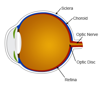
A brief discussion of the function of the retina will help in understanding PRA and its various forms. The retina is the innermost layer of the back of the eye, and it contains the cells that are sensitive to light ("photoreceptors") and the nerves structures that run from those cells through the optic nerve and, ultimately, to the brain. There are two types of photoreceptors: rods, which function in dim light, detect shape and motion, do not differentiate color, and are more concentrated around the perimeter of the retina; and cones, which function in brighter light, detect color, and are more concentrated in the central area of the retina.
The two major categories of retinal diseases typically grouped under Progressive Retinal Atrophy are (1) retinal dysplasia, where the key cells of the retina do not develop properly in the first eight weeks of life, and (2) retinal degeneration, where the cells do develop normally in the fetus and early puppyhood, but degenerate later in life. Additionally, some forms of PRA may affect only the rod cells or only the cone cells of the retina.
Link to this article... copy the code below and paste on your website
Symptoms of PRA
Although the types and expressions of the forms of Progressive Retinal Atrophy do vary in severity and age of onset, the common symptoms are progressive vision loss, almost always beginning with a loss of night vision, as the disease affects the rods first. As night vision deteriorates, the dog will typically exhibit behavioral signs, such as disorientation at night, apprehension around stairs or new environments in dim light, or literally getting lost in their own home, especially if the furniture has been rearranged. Physiologically, affected dogs will typically have excessively dilated pupils at night, as their eyes are trying to let in more light. Additionally, there may be a noticeably greater shine to the eyes at night, as the pupils dilate excessively and the retinal tissue thins, exposing more of the reflective rear surface of the eye.
Diagnosis of Progressive Retinal AtrophyPreliminary diagnosis is typically via fundoscopic exam (with an ophthalmoscope) by a licensed veterinary ophthalmologist. This exam is painless and requires no sedation, but the pupil must be dilated. The vet would be looking for blood vessel shrinkage, increased reflectivity of the tissue behind the retina, decreased pigmentation of the retina, and a darkened optic disc in advanced stages. However, secondary cataract formation, which may occur with the disease, may make examination difficult. Additionally, depending on the breed and type of Progressive Retinal Atrophy, these symptomatic changes may not occur until later in life when the disease begins to manifest, so a clear exam in a puppy does not guarantee they are free from the disease.
Often, though, a dog will already have exhibited signs of the disease, like night vision loss, by the time a fundoscopic exam would clearly identify PRA.
However, a more accurate assessment can be made via an electroretinogram (ERG), which is an electrical evaluation of the response of the retina to varying flashes of light and color. This procedure does require sedation, as it involves placing a type of contact lens over the eye. This procedure is sensitive and accurate enough to detect changes in the retina, often before the vision has deteriorated to the point that the dog shows signs of Progressive Retinal Atrophy.
Treatment of Progressive Retinal AtrophyThere is no treatment for PRA, and no known way to slow the progression. Generally, it eventually progresses to blindness. There have been some studies on the use of Vitamin A therapy on the human parallel disease, retinitis pigmentosa, but no studies have been done to date on vitamin therapy in dogs.
Breeds Affected by PRAFollowing are the breeds affected, type of PRA common within the breed, age of onset, genetic mode of inheritance, and presence of a genetic marker test for the breed.
| Breed | Type of PRA | Age of Onset | Inheritance | Genetic Test? |
|---|
| Akita | rod-cone degeneration | 1-3 yrs | autosomal recessive | No |
| American Eskimo Dog | rod-cone degeneration | 1-3 yrs | autosomal recessive | Yes |
| Bullmastiff | Rhodopsin deficiency | variable | autosomal dominant | Yes |
| Cairn Terrier | rod-cone dysplasia | < 1 yr | autosomal recessive | No |
| Cocker Spaniel | rod-cone degeneration | 3-6 yrs | autosomal recessive | Yes |
| Collie | rod-cone dysplasia | < 1 yr | autosomal recessive | No |
| Irish Setter | rod-cone dysplasia | < 1 yr | autosomal recessive | Yes |
| Labrador Retriever | rod-cone degeneration | 4-6 yrs | autosomal recessive | Yes |
| Min. LH Dachshund | generalized PRA | 6 mos | autosomal recessive | No |
| Miniature Poodle | rod-code degeneration | 3-6 yrs | autosomal recessive | Yes |
| Miniature Schnauzer | rod-code degeneration | 3-6 yrs | autosomal recessive | Yes |
| Norwegian Elkhound | early retinal degeneration | 6 wks – 1 yr | autosomal recessive | Yes |
| Papillon | generalized PRA | variable | autosomal recessive | No |
| Samoyed | X-Linked PRA | 3-5 yrs | X-Linked | Yes |
| Siberian Husky | X-Linked PRA | 2-4 yrs | X-linked | Yes |
| Tibetan Spaniel | generalized PRA | 3-5 yrs | autosomal recessive | No |
| Tibetan Terrier | generalized PRA | 6-12 mos | autosomal recessive | No |
Breeding DecisionsDogs affected with PRA should obviously not be allowed to breed, and should be neutered or spayed. Since Progressive Retinal Atrophy is inherited genetically, parents and siblings of PRA-affected dogs should also not be bred, in the absence of complete genetic evaluation. Fortunately, with precise genetic tests now available for many breeds, affected and carrier dogs can be pinpointed prior to breeding decisions.