Collie Eye Anomaly (CEA)
Collie Eye Anomaly (CEA) is an inherited disorder where tissues in the back of the eye do not differentiate and develop properly in the fetal puppy. The result is a group of defects which occur in varying degrees among different dogs, and even varying degrees between the right and left eyes of the same dog.
The primary defect is choroidal hypoplasia, where the choroid is under-developed. The choroid is a thin layer of blood vessels, sandwiched between the retina (most inner layer) and sclera (outer layer) of the back of the eye. The choroid supplies the retina with oxygen and nutrients, so when it is under-developed, the back tissues of the eye are receiving reduced levels of oxygen and nutrients.
Coloboma of the retina and/or optic disc may also be present in Collie Eye Anomaly. A coloboma is a hole or fissure in the tissue, typically caused by a failure of the embryonic tissue to close properly.
Staphyloma is also commonly associated with CEA. A staphyloma is a thinning in the sclera, which is the outer layer of the back of the eye.
Perhaps most significantly, retinal detachment, either complete or partial, is associated with CEA. Retinal detachment may or may not produce bleeding, but it is the possible result of the other CEA issues, and retinal detachment is the major contributor to the partial or complete blindness that can result from Collie Eye Anomaly.
Link to this article... copy the code below and paste on your website
Diagnosis of Collie Eye Anomaly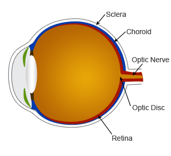
CEA can be clinically diagnosed by fundoscopic eye exam with dilation, as early as 5 to 6 weeks of age. In fact, early exam is highly recommended, as some retinal lesions become harder to detect as the puppy gets older. Since the condition is almost exclusive to the Collie breeds, but is not uncommon within those breeds, breeders are strongly advised to have their puppies checked at 5-6 weeks by a licensed veterinary ophthalmologist.
The most accurate information, though, will be via genetic testing. Optigen now offers a genetic test for the CEA gene, available for the collie breeds (genetic tests for diseases are often breed-specific). This test can identify both affected individuals and carriers, and is therefore critical in breeding decisions for Collie-breed breeders.
Breeds AffectedAs the name implies, Collie Eye Anomaly primarily affects the collie breeds, including the Rough and Smooth Collie varieties, Border Collie, and Shetland Sheepdog. Although it is rare, it has been found in the Borzoi, Beagle, Miniature Poodle, Toy Poodle, and Nova Scotia Duck Tolling Retriver.
How CEA Is InheritedCollie Eye Anomaly is an autosomal recessive trait, which means that both parents must carry the gene, either as affected individuals or as carriers. Since it is simple recessive, carriers will not show any symptoms, but two carriers bred together will produce, on average, 25% affected puppies, 50% carrier puppies, and 25% normal puppies. Therefore, the development of the Optigen genetic test that can identify carriers has been critical in the effort to eliminate this condition from the affected breeds.
Treatment and PrognosisThere is no treatment for Collie Eye Anomaly. The extent to which it causes vision problems varies, with the worst outcome being complete retinal detachment and total blindness, either in one or both eyes. This does typically occur before 2 years of age.